facs buffer flow cytometry
Cells are incubated with patient serum and an AlexaFluor 647-labeled secondary antibody is added. MOG-IgG1 Fluorescence-Activated Cell Sorting Assay FACS Human embryonic kidney cells.
Flow Cytometry And Cell Sorting By Facs In The Flow Cell 1 The Download Scientific Diagram
Add 100 μl of Fc block to each sample Fc block diluted in FACS buffer at 150 ratio.

. Flow cytometry multicolor experiments may need compensation when there is fluorescence spillover Figure 1Pairing fluorochromes based on antigen density fluorochrome brightness and separating by channels helps to minimize the effects from spillover and may remove the need for compensation from smaller experiments. BD FACSelect Buffer Compatibility Tool. If you are unable to immediately read your samples on a cytometer keep them shielded from light and in a refrigerator set at 4-8C.
Fluorophore and reagent selection guide for flow cytometry. Spectral Flow Cytometry Fundamentals. Incubate for at least 30 min at room temperature or 4C in.
A buffer system with different pH values is applied in gel electrophoresis process. If MOG-IgG1 cell based flow cytometry FACS assay is positive at screening dilution the MOG. People use protein containing buffers for flow cytometry is to prevent cells from sticking to the side of plastic tubes or other culture labware as well as preventing cell clumping.
However as the number of parameters and colors. BD FACS Sample Prep Assistant SPA III. Are lifted and resuspended in live cell-binding buffer.
These guidelines are a consensus work of a considerable number of members of the immunology and flow cytometry community. However they can be stained in any container for which you have an. Cells are usually stained in polystyrene round bottom 12 x 75 mm 2 Falcon tubes.
When gating on cell populations the light scatter profiles of the cells on the flow cytometer will change considerably after permeabilization. General procedure for flow cytometry using a conjugated primary antibody. Download Flow Cytometry Protocols Handbook.
Flow Cytometry Staining Buffer Catalog FC001 or an equivalent solution containing BSA and sodium azide. For more technical resources on flow cytometry visit. 1- Use CaMg2 free PBS.
Centrifuge at 1500 rpm for 5 min at 4C. 10 μgmL PI in PBS store at 4 C in the dark Materials Required. Detecting intracellular antigens requires cell permeabilization before staining.
Our flow cytometry expertise world class support and cutting-edge technology enable the routine or complex fluorescence activated cell sorting FACS analysis needed to propel your labs goals. BD FACSDuet Sample Preparation System. A debris free single cell suspension will have lower auto-fluorescence and flow smoothly through the nozzle.
Incubate on ice for 20 min. Dilutions if necessary should be made in FACS buffer. We use a dual labeling strategy to simplify the staining scheme and show the distribution of major peripheral blood lineages myeloid B cells T cells.
Add 01-10 μgml of the primary labeled antibody. FACS Tubes 5 mL round-bottom polystyrene tubes Pipette Tips and Pipettes. The samples should be resuspended in Cell Staining Buffer.
For more than 45 years we at BD Biosciences have dedicated ourselves to advancing science and improving lives through flow cytometry. Be part of the flow cytometry community with the latest flow cytometry news thought leader opinions blogs on breakthrough research. Antibodies should be prepared in permeabilization buffer to ensure the cells remain permeable.
Absence of these ions reduces cation-dependent cell to cell adhesion and prevents clumping. Here are 5 ingredients to consider for your FACS buffer. A very widespread discontinuous buffer system is the tris-glycine or Laemmli system that stacks at a pH of 68 and resolves at a pH of 83-90.
Invitrogen eBioscience ResourcesSelection guides Best Protocols product performance and more. With the development of more fluorochromes for flow cytometry it is now possible to analyze engraftment of the test cell population and multiple peripheral blood lineages with a single tube. They provide the theory and key practical aspects of flow cytometry enabling immunologists to avoid the common errors that often.
View All Reagent Selection Tools. Harvest wash the cells and adjust cell suspension to a concentration of 1-5 x 10 6 cellsmL in ice-cold PBS 10 FCS 1 sodium azide. Perform fluorescence activated cell sorting FACS or flow cytometric analysis.
Intracellular Staining for Flow Cytometry How-To Videofor detecting cytokines and intranuclear markers.

Flow Cytometry Gating Strategy For Hiv 1tg And F344 Rat Blood Download Scientific Diagram

Gating Strategy For Flow Cytometry A Strategy For Hek 293 Cells B Download Scientific Diagram

Facs Flow Cytometry Technical Blogs Bosterbio

Fluorescence Activated Cell Sorting Facs Sino Biological

Gating Strategy Used Throughout The Experiments For Flow Cytometry Download Scientific Diagram

Flow Cytometry And Cell Sorting By Facs In The Flow Cell 1 The Download Scientific Diagram

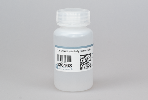
Flow Cytometry Antibody Dilution Buffer Cell Signaling Technology

Flow Cytometry Not As Scary As It Sounds A Bit Of Housekeeping About Me About You Institute Prior Knowledge Core Facility Ppt Download

Flow Cytometric Characterization Of Tissue Resident Lymphocytes After Murine Liver And Heart Transplantation Star Protocols

Flow Cytometry Sample Preparation

Functional Flow Cytometry Of Monocytes For Routine Diagnosis Of Innate Primary Immunodeficiencies Journal Of Allergy And Clinical Immunology

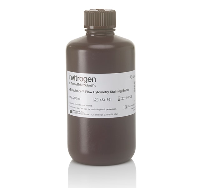
Ebioscience Flow Cytometry Staining Buffer

Flow Cytometry Facs Protocols Sino Biological

Buffers For Facs Analysis Download Table

Flow Cytometry Not As Scary As It Sounds A Bit Of Housekeeping About Me About You Institute Prior Knowledge Core Facility Ppt Download

Flow Cytometry Perm Buffer 10x Pf00011 C Proteintech

Gating Strategy Used Throughout The Experiments For Flow Cytometry Download Scientific Diagram

